|
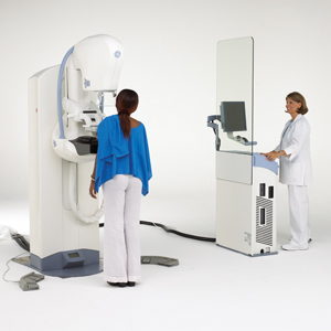
 Digital Mammography with Computer-Aided Detection (CAD) is a state of the art mammography system in which x-ray is replaced with solid-state detectors and computers. The specialized mammography system allows for short examination time and less radiation than traditional x-ray mammograms. Images are digitized allowing the radiologist more manipulation of images to further magnify, invert and enhance the picture for a more thorough evaluation of the breast improving detection of small tumors. CAD scans the digital image and automatically flags potentially suspicious area, alerting the radiologist to take a second look. The use of CAD may help to improve the accuracy of screening mammography resulting in early detection. Digital Mammography with Computer-Aided Detection (CAD) is a state of the art mammography system in which x-ray is replaced with solid-state detectors and computers. The specialized mammography system allows for short examination time and less radiation than traditional x-ray mammograms. Images are digitized allowing the radiologist more manipulation of images to further magnify, invert and enhance the picture for a more thorough evaluation of the breast improving detection of small tumors. CAD scans the digital image and automatically flags potentially suspicious area, alerting the radiologist to take a second look. The use of CAD may help to improve the accuracy of screening mammography resulting in early detection.
Screening Mammogram is a radiologic procedure provided to a woman without signs or symptoms of the breast for the purpose of early detection of breast cancer.
Diagnostic Mammogram is a radiologic procedure provided to a man or woman with signs and symptoms of the breast, further testing from an abnormal screening or if a patient has a personal history of breast cancer.
Ultrasound is an imaging technique for diagnosing breast symptoms. It uses harmless, high frequency sound waves to form an image (sonogram). The sound waves pass through the breast and bounce back or echo from various tissues to form a picture of the internal structures. It is not invasive and involves no radiation.
US Guided Cyst Aspiration is a simple procedure performed by placing an ultrasound probe over the site of a breast cyst and numbing the area with local anesthesia. The breast radiologist then places a small needle directly into the cyst and withdraws fluid.
Us Guided Core Biopsy is performed by a breast radiologist using an ultrasound probe to visualize the location of the breast mass, distortion or abnormal tissue change. The radiologist inserts a biopsy needle through the skin, advances it into the targeted finding and removes tissue samples. Ultrasound is used to guide a clip directly into the targeted finding to mark the area of the biopsy site for future referencing.
|